Abdominal Ultrasound
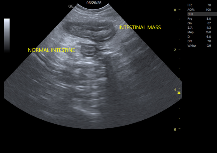
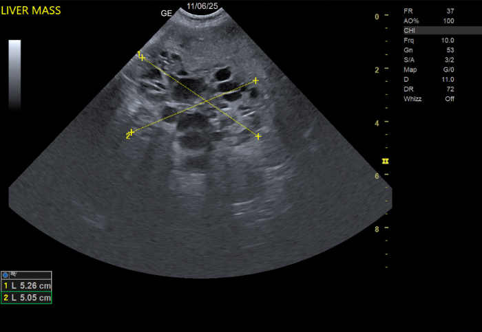
Ultrasound imaging, also known as diagnostic ultrasonography, is a safe, non-invasive tool that veterinarians use to evaluate your pet’s internal organs in real time. By using high-frequency sound waves, ultrasound creates detailed images of soft tissues such as the liver, kidneys, bladder, heart, and abdominal structures—without the need for radiation.
What Ultrasound is Used For
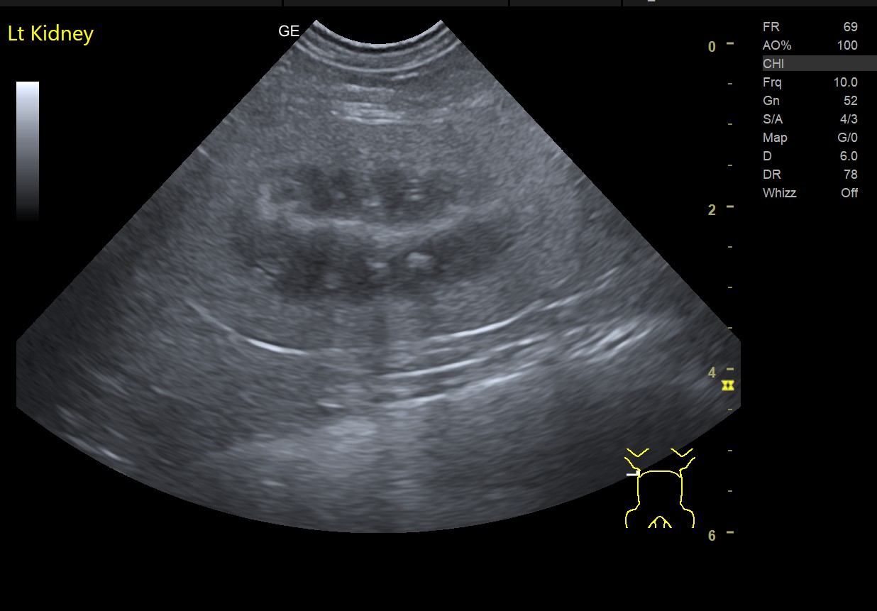
Ultrasound is especially helpful for examining soft tissues and fluid-filled structures. Common uses include:
- Evaluating abdominal organs such as the liver, kidneys, spleen and bladder
- Investigating the causes of vomiting, diarrhea, or weight loss
- Assessing the bladder and urinary tract for stones, tumors and cystitis
- Detecting pregnancy and monitoring fetal development
- Identifying tumors, cysts, and internal bleeding or fluid in the abdomen
What to Expect During the Procedure
Most pets are awake during an ultrasound. The area being examined may be shaved to improve image quality, and a special gel is applied to help transmit sound waves. In some cases, mild sedation may be recommended if your pet is anxious or uncomfortable.
Benefits of Ultrasound for Pets
- Non-invasive and painless
- No radiation exposure
- Real-time imaging for accurate diagnosis
- Helps guide procedures such as biopsies or fluid collection
When Is Ultrasound Recommended?
Your veterinarian may suggest an ultrasound if your pet shows symptoms like vomiting, unexplained weight loss, abdominal pain, or changes in urination. It is also commonly used as a follow-up to X-rays or bloodwork for a more detailed evaluation.
Ultrasound imaging is a valuable diagnostic tool that helps veterinarians detect problems early and develop effective treatment plans—keeping your pet healthier and happier.